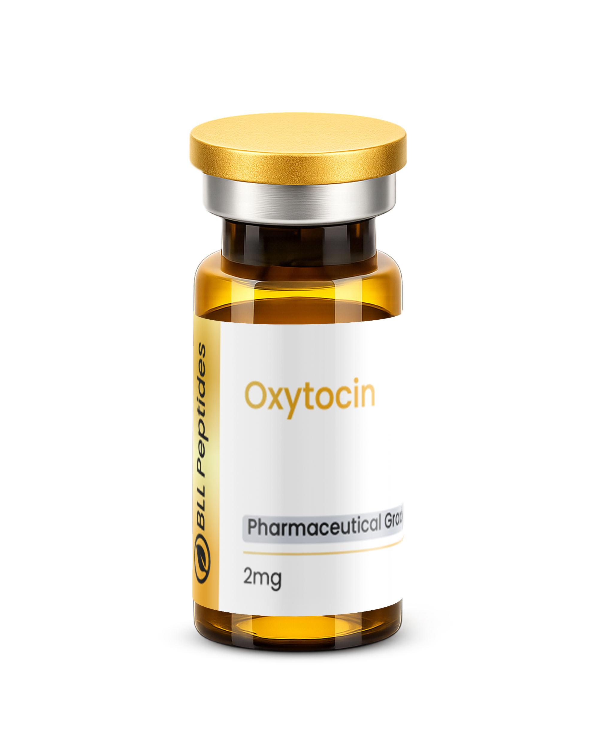
Oxytocin 2mg (3ml)
Oxytocin is a research-grade neuropeptide studied for roles in social bonding, trust modulation, stress response, maternal behavior, and cardiovascular regulation as a key neuroendocrine messenger. Researchers investigating neuroendocrine function, behavioral neuroscience, and social cognition rely on pharmaceutical-grade purity for reliable experimental results. Available at BLL Peptides — USA-made, rigorously tested. ✅ COA tested every batch✅…
Description
Oxytocin: Complete Research Guide – Neuropeptide Hormone Mechanisms, Social Behavior Research, and Neuroendocrine Applications
Last updated: March 2026
Executive Summary
Oxytocin is a cyclic nonapeptide neurohormone composed of nine amino acids (Cys-Tyr-Ile-Gln-Asn-Cys-Pro-Leu-Gly-NH2) with a molecular formula of C43H66N12O12S2 and a molecular weight of approximately 1,007.19 Daltons (CAS: 50-56-6). Its defining structural feature is an intramolecular disulfide bridge between Cys1 and Cys6, which constrains the first six residues into a cyclic ring while the remaining three residues (Pro7-Leu8-Gly9-NH2) extend as a flexible C-terminal tail. This tocin ring-plus-tail architecture is shared with the structurally related neurohypophyseal hormone vasopressin, from which oxytocin differs at only two amino acid positions [1].
Oxytocin holds a singular place in the history of biochemistry as the first peptide hormone to have its structure determined and subsequently synthesized in the laboratory. Vincent du Vigneaud accomplished the total chemical synthesis of oxytocin in 1953, an achievement for which he was awarded the Nobel Prize in Chemistry in 1955. This landmark synthesis demonstrated that a biologically active hormone could be produced entirely from its constituent amino acids, establishing a foundational principle of modern peptide chemistry and neuroendocrinology [2].
In mammalian physiology, oxytocin is primarily synthesized by magnocellular neurosecretory neurons in the paraventricular nucleus (PVN) and supraoptic nucleus (SON) of the hypothalamus. These neurons project their axons to the posterior pituitary gland (neurohypophysis), where oxytocin is stored in dense-core vesicles and released into systemic circulation in response to specific physiological stimuli including cervical distension during parturition, nipple stimulation during lactation, and various social-tactile stimuli. Simultaneously, oxytocin is released centrally from dendrites and axon collaterals to act as a neuromodulator within the brain itself, influencing neural circuits involved in social cognition, pair bonding, anxiety regulation, and stress responsiveness [3, 4].
The biological effects of oxytocin are mediated through the oxytocin receptor (OTR), a Gq/11-coupled G-protein coupled receptor (GPCR) encoded by the OXTR gene on chromosome 3p25. OTR activation triggers phospholipase C-mediated hydrolysis of phosphatidylinositol 4,5-bisphosphate (PIP2) into inositol trisphosphate (IP3) and diacylglycerol (DAG), resulting in intracellular calcium mobilization and downstream signaling cascades. The distribution of OTR across uterine myometrium, mammary gland myoepithelial cells, and numerous brain regions underlies the remarkable diversity of oxytocin's physiological functions — from the powerful uterotonic contractions of labor to the subtle modulation of social trust and interpersonal bonding [5].
Clinically, synthetic oxytocin (marketed as Pitocin and Syntocinon) is one of the most widely administered medications in obstetric practice, with FDA approval for the induction and augmentation of labor as well as the management of postpartum hemorrhage. Beyond its established obstetric applications, oxytocin has become a major focus of translational research in neuropsychiatry, with intranasal administration studies investigating its potential effects on social cognition deficits in autism spectrum disorder (ASD), anxiety disorders, post-traumatic stress disorder (PTSD), and schizophrenia. The compound's dual identity — as both an essential reproductive hormone and a central modulator of complex social behaviors — has made it one of the most intensively studied neuropeptides in contemporary neuroscience [6, 7].
Interactive 3D Molecular Structure
The following interactive 3D visualization renders the oxytocin cyclic nonapeptide structure. The six-residue ring formed by the Cys1-Cys6 disulfide bridge is displayed as the core cyclic motif, with the three-residue C-terminal tail (Pro7-Leu8-Gly9) extending outward. The disulfide bond between the two cysteine residues is shown as a gold dashed line.
Legend: The interactive visualization above depicts the cyclic nonapeptide structure of oxytocin. The six-residue tocin ring — Cys1-Tyr2-Ile3-Gln4-Asn5-Cys6 — is closed by a disulfide bond (dashed gold line) between the two cysteine residues. The C-terminal tripeptide tail (Pro7-Leu8-Gly9-NH2) extends outward from Cys6. Gold nodes represent cysteine and polar residues (Cys, Gln, Asn), the teal node denotes the aromatic residue (Tyr), gray nodes indicate hydrophobic residues (Ile, Pro, Leu), and the light gray node marks the small residue (Gly). Drag to rotate; scroll to zoom.
Table of Contents
- Introduction and Development History
- Molecular Structure and Chemistry
- Detailed Mechanism of Action
- Scientific Research Review
- Comparison with Related Neuropeptides and Receptor Modulators
- Safety Profile and Pharmacology
- Research Applications
- References
- Disclaimer
Introduction and Development History
Discovery of Oxytocin and Early Neuroendocrinology
The history of oxytocin begins at the dawn of modern endocrinology. In 1906, Sir Henry Hallett Dale observed that extracts from the posterior pituitary gland caused powerful contractions of the uterus in pregnant cats, coining the term "oxytocin" from the Greek words οξυς (oxys, "swift") and τοκος (tokos, "birth") — literally meaning "quick birth." This observation established the existence of a uterotonic substance in the neurohypophysis and initiated nearly five decades of research aimed at isolating and characterizing the active principle [8].
In the 1920s and 1930s, Oliver Kamm and colleagues at Parke-Davis achieved a partial separation of the two major active principles of posterior pituitary extracts: one with pressor (vasopressin) and one with oxytocic activity. However, the complete isolation and structural characterization of oxytocin would require the development of more sophisticated chromatographic and analytical techniques. In 1950, Vincent du Vigneaud and colleagues at Cornell University Medical College began a systematic effort to isolate and determine the structure of the posterior pituitary hormones. Using countercurrent distribution and amino acid analysis, du Vigneaud's group established the complete amino acid sequence of oxytocin by 1953 as a cyclic nonapeptide with the sequence Cys-Tyr-Ile-Gln-Asn-Cys-Pro-Leu-Gly-NH2, with a disulfide bridge between the two cysteine residues [2, 9].
The Nobel Prize Synthesis
The crowning achievement in oxytocin research came in 1953 when du Vigneaud, along with collaborators including Ressler, Swan, Roberts, and Katsoyannis, accomplished the total chemical synthesis of oxytocin and demonstrated that the synthetic product possessed full biological activity identical to the natural hormone. This synthesis — the first total synthesis of a biologically active polypeptide hormone — represented a watershed moment in biochemistry. It definitively proved that the biological activity of a peptide hormone resides solely in its chemical structure and does not require any mysterious "vital force" or post-translational modifications beyond the disulfide bridge. For this work, du Vigneaud was awarded the 1955 Nobel Prize in Chemistry, with the Nobel committee specifically citing "his work on biochemically important sulphur compounds, especially for the first synthesis of a polypeptide hormone" [2].
The synthesis proceeded through a stepwise approach involving protection group chemistry for the reactive side chains, formation of the individual peptide bonds through classical mixed anhydride and azide coupling methods, and final oxidative cyclization to form the Cys1-Cys6 disulfide bond. While primitive by today's standards of solid-phase peptide synthesis (SPPS), this work established the fundamental principles and chemical feasibility of peptide hormone synthesis that would enable the entire field of synthetic peptide therapeutics [10].
From Obstetric Hormone to Social Neuropeptide
For decades following its synthesis, oxytocin was studied primarily as a peripheral hormone with two well-characterized functions: stimulation of uterine smooth muscle contraction during parturition and stimulation of milk ejection from the mammary gland during lactation. The introduction of synthetic oxytocin (Pitocin) into clinical obstetric practice in the 1960s represented one of the earliest therapeutic applications of a synthetic peptide hormone and remains one of the most widely used interventions in modern obstetrics [11].
The transformation of oxytocin from a "simple" reproductive hormone into a central focus of social neuroscience research began in the 1970s and accelerated dramatically in the 1990s and 2000s. Key milestones in this conceptual revolution include:
- 1979: Pedersen and Prange demonstrate that intracerebroventricular (ICV) oxytocin infusion induces maternal behavior in virgin rats, establishing that oxytocin acts centrally to influence social behavior [12]
- 1992-1994: C. Sue Carter, Lowell Getz, and colleagues demonstrate that oxytocin is critical for pair bond formation in the socially monogamous prairie vole (Microtus ochrogaster), providing the first evidence linking oxytocin to pair bonding behavior [13]
- 1999: Thomas Insel and Larry Young establish that species differences in OTR distribution in the brain account for species differences in social bonding behavior, creating a neuroanatomical framework for understanding oxytocin's social functions [14]
- 2005: Michael Kosfeld, Markus Heinrichs, and colleagues publish the landmark "trust study" in Nature, demonstrating that intranasal oxytocin increases trust behavior in human subjects during an economic trust game, igniting widespread interest in oxytocin's role in human social cognition [15]
- 2010: Andari and colleagues publish the first controlled study of intranasal oxytocin in individuals with autism spectrum disorder, demonstrating improved social interaction during a ball-tossing game [16]
- 2012-present: Growing recognition of the complexity and context-dependence of oxytocin's social effects, with research demonstrating that oxytocin can enhance both prosocial and antisocial behaviors depending on the social context, individual characteristics, and group membership [17]
Key Historical Milestones
| Year | Milestone |
|---|---|
| 1906 | Henry Dale discovers oxytocic activity in posterior pituitary extracts |
| 1928 | Kamm separates oxytocic and vasopressor fractions from pituitary extract |
| 1950-1953 | du Vigneaud determines the amino acid sequence of oxytocin |
| 1953 | First total synthesis of oxytocin — first synthetic peptide hormone |
| 1955 | du Vigneaud awarded Nobel Prize in Chemistry |
| 1960s | Synthetic oxytocin (Pitocin) introduced into obstetric practice |
| 1979 | Central oxytocin shown to induce maternal behavior in rats |
| 1992 | Oxytocin linked to pair bonding in prairie voles |
| 2005 | Intranasal oxytocin shown to increase trust in humans |
| 2010 | First controlled oxytocin trial in autism spectrum disorder |
| 2019 | FDA approves Pitocin label expansion; ongoing neuropsychiatric research |
Molecular Structure and Chemistry
Amino Acid Sequence and the Tocin Ring
Oxytocin is a cyclic nonapeptide (nine amino acids) with the primary sequence:
H-Cys1-Tyr2-Ile3-Gln4-Asn5-Cys6-Pro7-Leu8-Gly9-NH2
The structural features include:
-
Tocin ring (residues 1-6): The intramolecular disulfide bond between the sulfhydryl groups of Cys1 and Cys6 creates a 20-atom ring (the "tocin ring") that constrains the backbone and side chains of the first six residues into a relatively rigid cyclic conformation. This ring is essential for receptor binding and biological activity; reduction of the disulfide bond abolishes oxytocic activity [1, 18]
-
C-terminal tripeptide tail (residues 7-9): The sequence Pro7-Leu8-Gly9-NH2 extends from Cys6 as a flexible, linear tail. The proline residue introduces a conformational kink due to its cyclic pyrrolidine side chain, while the C-terminal glycinamide (-Gly-NH2) is generated by amidation of the C-terminal carboxyl group. This amidation is critical for full biological activity; the free acid form (Gly-OH) exhibits dramatically reduced potency [18]
-
Tyrosine at position 2: Tyr2 is the sole aromatic residue in oxytocin and plays a critical role in receptor binding. Iodination or nitration of the Tyr2 phenolic ring modulates receptor affinity, and Tyr2 has been a key site for radioligand labeling (e.g., 125I-oxytocin) in receptor binding studies
-
Isoleucine at position 3: The presence of Ile3 distinguishes oxytocin from [Arg8]-vasopressin (AVP), which has Phe at position 3. This single substitution contributes to the differential receptor selectivity between the two neurohypophyseal hormones
Physicochemical Properties
| Property | Value |
|---|---|
| Molecular Formula | C43H66N12O12S2 |
| Molecular Weight | Approximately 1,007.19 Da |
| CAS Number | 50-56-6 |
| Appearance | White to off-white lyophilized powder |
| Solubility | Freely soluble in water; soluble in n-butanol and aqueous acetic acid |
| Peptide Type | Cyclic disulfide-bridged nonapeptide |
| Sequence Length | 9 amino acids (6-residue ring + 3-residue tail) |
| Cyclization | Cys1-Cys6 intramolecular disulfide bond |
| Isoelectric Point (pI) | Approximately 7.7 |
| Plasma Half-Life | Approximately 3-6 minutes (intravenous) |
| Storage | Lyophilized: -20°C; Reconstituted: 2-8°C, use within 28 days |
| pH Stability | Most stable at pH 3-5 in aqueous solution |
| Endogenous Source | Hypothalamic PVN and SON neurons; released from posterior pituitary |
Structural Comparison: Oxytocin vs. Vasopressin
Oxytocin and [Arg8]-vasopressin (AVP) are the two neurohypophyseal hormones in most placental mammals, sharing a common evolutionary ancestor and differing at only two of nine amino acid positions:
Oxytocin: Cys-Tyr-Ile-Gln-Asn-Cys-Pro-Leu-Gly-NH2
Vasopressin (AVP): Cys-Tyr-Phe-Gln-Asn-Cys-Pro-Arg-Gly-NH2
The two critical differences are:
- Position 3: Ile (oxytocin) vs. Phe (vasopressin) — aliphatic vs. aromatic hydrophobic side chain
- Position 8: Leu (oxytocin) vs. Arg (vasopressin) — hydrophobic vs. basic charged side chain
Despite these minimal sequence differences, the two peptides show remarkable selectivity for their respective receptors (OTR vs. V1a/V1b/V2 receptors), though significant cross-reactivity exists at high concentrations. The Arg8 residue in vasopressin is particularly important for V2 receptor selectivity and antidiuretic activity [18, 19].
Stability and Degradation
Oxytocin is susceptible to several degradation pathways:
- Enzymatic degradation: Oxytocinase (leucyl-cystinyl aminopeptidase, also known as placental leucine aminopeptidase or P-LAP, encoded by the LNPEP gene) is the primary degrading enzyme in circulation, cleaving the Cys1-Tyr2 bond after reduction of the disulfide bridge. During pregnancy, oxytocinase levels increase approximately 10-fold, providing a physiological mechanism for regulating circulating oxytocin levels [20]
- Disulfide exchange: The Cys1-Cys6 disulfide bond can undergo thiol-disulfide exchange in the presence of reducing agents or free thiol-containing compounds, resulting in ring opening and loss of activity
- Oxidation: The Tyr2 residue can undergo oxidation, and the methionine-free sequence of oxytocin provides inherent resistance to methionine oxidation (unlike vasopressin analogs)
- Deamidation: The Gln4 and Asn5 residues are susceptible to non-enzymatic deamidation under alkaline or elevated temperature conditions
These stability considerations are critical for research formulation, storage, and intranasal delivery development [1].
Detailed Mechanism of Action
Oxytocin Receptor (OTR) Structure and Distribution
Oxytocin exerts its biological effects primarily through the oxytocin receptor (OTR), a class A rhodopsin-family G-protein coupled receptor (GPCR) consisting of 389 amino acids arranged in the canonical seven-transmembrane domain architecture. The OXTR gene is located on chromosome 3p25.3 in humans and encodes a receptor with an extracellular N-terminal domain, seven transmembrane α-helices connected by three extracellular and three intracellular loops, and an intracellular C-terminal domain [5].
OTR is expressed in a wide variety of tissues:
Peripheral tissues:
- Uterine myometrium (dramatically upregulated during pregnancy, increasing approximately 300-fold by term)
- Mammary gland myoepithelial cells
- Ovarian corpus luteum
- Testicular Leydig cells
- Cardiac myocytes
- Endothelial cells
- Kidney
- Adipose tissue
Central nervous system:
- Hypothalamus (PVN, SON, ventromedial nucleus)
- Amygdala (central and medial nuclei)
- Hippocampus
- Nucleus accumbens and ventral pallidum
- Prefrontal cortex
- Brainstem nuclei (nucleus tractus solitarius, dorsal motor nucleus of vagus)
- Olfactory bulb
Signal Transduction Pathways
Upon oxytocin binding, OTR activates multiple intracellular signaling cascades:
Primary pathway — Gq/11 coupling:
- OTR activation stimulates Gq/11 proteins, which activate phospholipase C-β (PLC-β)
- PLC-β hydrolyzes PIP2 into IP3 and DAG
- IP3 binds to IP3 receptors on the endoplasmic reticulum, triggering intracellular Ca2+ release
- Elevated Ca2+ activates myosin light chain kinase (MLCK) in smooth muscle cells, leading to actin-myosin cross-bridge cycling and contraction (uterine contractions, milk ejection)
- DAG activates protein kinase C (PKC), which modulates diverse downstream targets including ion channels and transcription factors [5, 21]
Secondary pathways:
- Gi coupling: In some cell types, OTR can couple to Gi proteins, inhibiting adenylyl cyclase and reducing cAMP levels
- MAPK/ERK pathway: OTR activation triggers ERK1/2 phosphorylation through both G-protein-dependent and β-arrestin-dependent mechanisms, contributing to cell proliferation and differentiation effects
- eNOS activation: In endothelial cells, OTR stimulation increases endothelial nitric oxide synthase (eNOS) activity, promoting nitric oxide (NO) production and vasodilation
- AMPK pathway: Recent evidence indicates oxytocin activates AMP-activated protein kinase (AMPK) in cardiac and adipose tissue, potentially contributing to metabolic effects [22]
Peripheral Actions: Uterine Contraction and Lactation
The two classical peripheral actions of oxytocin are directly explained by OTR signaling:
Uterotonic activity: During pregnancy, uterine OTR expression increases progressively, reaching peak density at term. Oxytocin binding to myometrial OTR triggers rhythmic uterine contractions through the Gq/PLC/Ca2+/MLCK pathway. The sensitivity of the uterus to oxytocin increases approximately 100-fold during pregnancy due to combined effects of OTR upregulation (driven by rising estrogen levels) and gap junction formation between myometrial cells (mediated by connexin-43), enabling coordinated uterine contractility. This mechanism forms the physiological basis for the clinical use of synthetic oxytocin (Pitocin) for labor induction and augmentation [11, 21].
Milk ejection reflex: Suckling stimulation activates mechanoreceptors in the nipple, which send afferent signals through the spinothalamic tract to the hypothalamus, triggering burst firing of oxytocin-secreting neurons in the PVN and SON. The resulting pulse of oxytocin release into the circulation activates OTR on mammary gland myoepithelial cells, causing contraction and ejection of milk from the alveoli into the ductal system. This neuroendocrine reflex operates on a rapid timescale (approximately 30-60 seconds from suckling to milk ejection) and demonstrates the characteristic pulsatile secretion pattern of oxytocin [3].
Central Actions: Social Cognition and Behavioral Modulation
The central actions of oxytocin are mediated by OTR in brain regions involved in social cognition, emotion regulation, and reward processing. Key neural circuits include:
Social recognition and memory: Oxytocin signaling in the medial amygdala and hippocampus is critical for social recognition memory — the ability to identify and remember conspecifics. OTR knockout mice and oxytocin knockout mice display impaired social recognition despite intact non-social memory [14, 23].
Pair bonding and attachment: In prairie voles, oxytocin release during mating activates OTR in the nucleus accumbens and prefrontal cortex, facilitating the formation of selective partner preferences. The density and distribution of OTR in reward-related brain regions differs between monogamous and non-monogamous vole species, providing a neurobiological basis for species differences in social bonding behavior [13, 14].
Anxiolysis and stress modulation: Oxytocin acts in the central amygdala, bed nucleus of the stria terminalis (BNST), and PVN to attenuate the hypothalamic-pituitary-adrenal (HPA) axis stress response. Central oxytocin release reduces cortisol secretion, decreases amygdala activation in response to threatening social stimuli, and promotes approach behavior in social contexts. This anxiolytic action has been proposed as a key mechanism through which oxytocin facilitates social engagement [6, 15].
Reward processing: OTR expression in the ventral tegmental area (VTA) and nucleus accumbens positions oxytocin at the interface of social and reward circuitry. Oxytocin modulates dopaminergic signaling in mesolimbic pathways, potentially contributing to the rewarding aspects of social interaction and the motivation to seek social contact [24].
Scientific Research Review
Human Intranasal Oxytocin Studies: Social Cognition
The development of intranasal oxytocin (IN-OT) administration as a non-invasive method for delivering oxytocin to the brain has enabled a large body of human behavioral research. While the precise pharmacokinetics and brain penetrance of intranasally administered oxytocin remain debated, functional neuroimaging studies have consistently demonstrated that IN-OT modulates activity in brain regions expressing OTR [6].
Trust and economic decision-making: The landmark study by Kosfeld et al. (2005) demonstrated that IN-OT (24 IU) significantly increased the amount of money participants entrusted to an anonymous partner in a trust game, compared to placebo. Critically, this effect was specific to social risk-taking; oxytocin did not increase risk-taking in a non-social gambling task, suggesting selective modulation of social cognition rather than general risk tolerance. Subsequent studies by Baumgartner et al. (2008) showed that oxytocin maintained trust even after explicit betrayal, implicating altered processing of negative social feedback [15, 25].
Emotion recognition and empathy: Multiple studies have demonstrated that IN-OT enhances the recognition of subtle emotional expressions, particularly in the eye region of the face. Domes et al. (2007) reported that oxytocin specifically improved the recognition of emotional states from photographs of the eye region using the "Reading the Mind in the Eyes" test (RMET). This finding is particularly relevant to autism research, as individuals with ASD frequently show deficits in this task [26].
Social memory: Rimmele et al. (2009) demonstrated that IN-OT selectively enhanced recognition memory for faces but not for non-social visual stimuli, consistent with a preferential effect on social information processing. This social specificity has been replicated across multiple laboratories and experimental paradigms [27].
Autism Spectrum Disorder Research
Oxytocin has been investigated as a potential therapeutic for the social communication deficits characteristic of ASD, based on converging evidence including reduced peripheral oxytocin levels in some ASD populations, genetic associations between OXTR variants and autism risk, and animal model data demonstrating that oxytocin system disruption impairs social behavior [7].
Single-dose studies: Andari et al. (2010) found that a single intranasal dose of oxytocin (24 IU) improved social interaction behavior in adults with high-functioning ASD during a computerized ball-tossing game, increasing their attention to socially relevant partners. Neuroimaging studies by Domes et al. (2013) demonstrated that IN-OT normalized amygdala and fusiform gyrus activation during face processing in individuals with ASD [16].
Chronic administration trials: Longer-term studies have produced more mixed results. Yatawara et al. (2016) conducted a systematic review and meta-analysis of oxytocin trials in ASD, finding modest but statistically significant improvements in some measures of social cognition after repeated administration, though effect sizes were generally small and clinical significance uncertain. The large-scale SOAR (Study of Oxytocin in Autism to improve Reciprocal social behaviors) trial, published by Sikich et al. (2021) in the New England Journal of Medicine, enrolled 290 children and adolescents with ASD in a 24-week randomized, placebo-controlled trial of intranasal oxytocin. The trial found no significant improvement in social functioning on the primary outcome measure (the Aberrant Behavior Checklist-Modified Social Withdrawal subscale), tempering enthusiasm for oxytocin as a standalone ASD treatment while highlighting the need for more targeted approaches [7, 28].
Anxiety and Stress-Related Disorders
The anxiolytic properties of oxytocin have been investigated in both non-clinical populations and individuals with anxiety disorders:
Social anxiety disorder: Labuschagne et al. (2010) demonstrated that IN-OT (24 IU) reduced amygdala hyperactivity in response to fearful faces in individuals with generalized social anxiety disorder using functional magnetic resonance imaging (fMRI). Subsequent studies by Guastella et al. (2009) showed that IN-OT enhanced the benefit of exposure therapy for social anxiety when administered prior to therapy sessions, suggesting potential utility as an adjunct to psychotherapy [29].
Post-traumatic stress disorder (PTSD): Preclinical studies demonstrate that oxytocin in the amygdala facilitates fear extinction — the process by which conditioned fear responses are weakened through repeated exposure to the conditioned stimulus without the unconditioned stimulus. In human PTSD research, van Zuiden et al. (2017) conducted a randomized trial of repeated intranasal oxytocin administration starting within 12 days of trauma exposure in emergency department patients, finding that oxytocin reduced the development of PTSD symptoms at 1.5 months follow-up. However, the effect was modulated by individual differences, with greater benefit in participants with higher acute distress levels [30].
HPA axis regulation: Heinrichs et al. (2003) provided foundational evidence that IN-OT combined with social support additively reduced cortisol responses to the Trier Social Stress Test (TSST) in healthy men. This combined social-neuroendocrine buffering effect has been replicated and extended to demonstrate that oxytocin enhances the stress-buffering effects of social support across diverse stress paradigms [6].
Pair Bonding and Attachment Research
Prairie vole model: The prairie vole has served as the principal animal model for understanding oxytocin's role in pair bonding. Williams et al. (1994) demonstrated that intracerebroventricular oxytocin infusion facilitated partner preference formation in female prairie voles without mating, while OTR antagonist infusion prevented mating-induced partner preference formation. Young et al. (2001) showed that viral vector-mediated overexpression of OTR in the ventral pallidum of non-monogamous meadow voles was sufficient to induce partner preference formation, establishing a direct causal link between OTR distribution and bonding behavior [13, 14].
Human attachment: Schneiderman et al. (2012) reported that plasma oxytocin levels were elevated in the early stages of romantic attachment (first 3 months) compared to singles and were correlated with interactive reciprocity between partners. Feldman et al. (2012) demonstrated that higher salivary oxytocin levels in new parents were associated with greater parent-infant synchrony, including more affectionate touch, gaze, and vocal affect during interactions [31].
Wound Healing and Regenerative Research
Emerging research has identified oxytocin as a modulator of tissue repair and regeneration. Elabd et al. (2014) demonstrated that oxytocin signaling declines with age in mice and that systemic oxytocin administration restored muscle stem cell (satellite cell) activation and muscle regeneration capacity in aged mice. OTR was found to be expressed on muscle stem cells, and oxytocin treatment activated MAPK/ERK signaling to promote myogenic differentiation. These findings suggest that age-related decline in oxytocin may contribute to sarcopenia and impaired tissue repair, positioning oxytocin as a potential factor in regenerative biology research [32].
Comparison with Related Neuropeptides and Receptor Modulators
Oxytocin vs. Vasopressin
| Parameter | Oxytocin | Vasopressin (AVP) |
|---|---|---|
| Sequence | Cys-Tyr-Ile-Gln-Asn-Cys-Pro-Leu-Gly-NH2 | Cys-Tyr-Phe-Gln-Asn-Cys-Pro-Arg-Gly-NH2 |
| Molecular Weight | Approximately 1,007.19 Da | Approximately 1,084.23 Da |
| Primary Receptor | OTR (Gq/11-coupled) | V1aR, V1bR (Gq), V2R (Gs) |
| Hypothalamic Source | PVN (primarily), SON | PVN, SON |
| Key Peripheral Actions | Uterine contraction, milk ejection | Vasoconstriction, water reabsorption |
| Key Central Actions | Social bonding, anxiolysis, trust | Aggression regulation, social memory, stress |
| Cross-Reactivity | Moderate affinity for V1a/V1b at high concentrations | Low-moderate affinity for OTR |
| Plasma Half-Life | Approximately 3-6 minutes | Approximately 10-20 minutes |
| Clinical Use | Labor induction (Pitocin) | Diabetes insipidus, variceal bleeding |
Oxytocin vs. Carbetocin (Long-Acting Analog)
| Parameter | Oxytocin | Carbetocin |
|---|---|---|
| Structure | Native cyclic nonapeptide | Deamino-1-monocarba-(2-O-methyltyrosine)-oxytocin |
| Half-Life | Approximately 3-6 minutes (IV) | Approximately 40 minutes (IV) |
| Modifications | None (native sequence) | Deamination of Cys1, methylation of Tyr2, thioether replacing disulfide |
| Receptor Selectivity | OTR agonist with some V1a/V1b activity | OTR partial agonist, improved selectivity |
| Clinical Use | Labor induction, postpartum hemorrhage | Prevention of postpartum hemorrhage (WHO Essential Medicine) |
| Thermostability | Requires cold chain storage | Heat-stable formulation available (critical for resource-limited settings) |
| Route | IV, IM, intranasal | IV, IM |
Oxytocin Research Context with PT-141
While oxytocin and PT-141 (bremelanotide) operate through entirely different receptor systems (OTR vs. melanocortin-4 receptor), both peptides modulate aspects of social and reproductive behavior through central nervous system mechanisms. Oxytocin influences the affiliative and bonding dimensions of social behavior through hypothalamic and limbic circuits, while PT-141 targets sexual motivation through melanocortin signaling in overlapping hypothalamic regions. Research has identified functional interactions between the oxytocin and melanocortin systems in the hypothalamus, with oxytocin neurons in the PVN expressing MC4R and responding to melanocortin agonists, suggesting potential synergistic or modulatory relationships between these neuropeptide systems in the regulation of social-reproductive behavior [33].
Comparison of Oxytocin Administration Routes
| Parameter | Intravenous (IV) | Intramuscular (IM) | Intranasal (IN) |
|---|---|---|---|
| Onset | 1-3 minutes | 3-5 minutes | 15-30 minutes (central effects) |
| Peak Effect | Immediate (continuous infusion) | 30-60 minutes | 30-60 minutes |
| Duration | Minutes after stopping infusion | 30-60 minutes | 2-4 hours (behavioral effects) |
| Primary Use | Labor induction/augmentation | Postpartum hemorrhage prevention | Research (social behavior studies) |
| Bioavailability | 100% | High | Approximately 2-5% (systemic); central delivery debated |
| CNS Penetrance | Minimal (poor BBB crossing) | Minimal | Proposed nose-to-brain pathway via olfactory/trigeminal nerves |
| FDA Status | Approved (Pitocin) | Approved | Research use only (for behavioral indications) |
Safety Profile and Pharmacology
Pharmacokinetics
Intravenous administration: Oxytocin administered intravenously has a plasma elimination half-life of approximately 3-6 minutes, with steady-state levels achieved rapidly during continuous infusion. The volume of distribution is approximately 12.2 liters, and clearance is approximately 20 mL/min/kg. Oxytocin is primarily metabolized by oxytocinase (leucyl-cystinyl aminopeptidase) in the plasma and by the liver and kidneys. Less than 1% of the administered dose is excreted unchanged in the urine [11, 20].
Intranasal administration: The pharmacokinetics of intranasally administered oxytocin are more complex and less well-characterized. Following IN-OT administration (typically 24-40 IU), plasma oxytocin levels rise within 10-15 minutes and peak at approximately 30-60 minutes, with elevations persisting for 60-120 minutes. However, the relationship between peripheral plasma levels and central nervous system concentrations is unclear. Striepens et al. (2013) reported that intranasal oxytocin elevated cerebrospinal fluid (CSF) oxytocin concentrations by approximately 2.5-fold at 75 minutes post-administration in humans, providing evidence for nose-to-brain transport, though the relative contributions of direct neural pathway transport versus peripheral-to-central feedback remain debated [34].
Known Adverse Effects (Clinical Oxytocin Use)
The safety profile of oxytocin is well-characterized in the context of obstetric use:
Common adverse effects:
- Uterine hyperstimulation (tachysystole) with IV infusion for labor induction
- Nausea and vomiting
- Headache
- Tachycardia or reflex bradycardia
Serious adverse effects (obstetric context):
- Water intoxication and hyponatremia (due to antidiuretic effects at high doses, mediated by cross-reactivity with V2 receptors)
- Uterine rupture (extremely rare, associated with excessive dosing or contraindicated use)
- Neonatal hyperbilirubinemia
- Arrhythmias (rare, associated with IV bolus administration)
Intranasal oxytocin (research context):
The safety profile of intranasal oxytocin in research studies has been generally favorable. MacDonald et al. (2011) conducted a comprehensive review of 38 randomized controlled trials involving IN-OT administration in human participants and found that adverse effects were comparable to placebo, with the most commonly reported events being mild nasal irritation, drowsiness, and headache. No serious adverse events were attributed to IN-OT in controlled research settings [35].
Drug Interactions and Contraindications
- Prostaglandins: Concurrent use with prostaglandin analogs (misoprostol, dinoprostone) may cause uterine hyperstimulation
- Vasoconstrictor medications: Combined with ergot alkaloids, oxytocin may increase the risk of hypertension
- Cyclopropane anesthesia: Historical concern for cardiovascular effects (hypotension followed by reflex tachycardia) when oxytocin is administered during cyclopropane anesthesia
- CYP3A4 considerations: Oxytocin is not significantly metabolized by CYP enzymes, minimizing hepatic drug-drug interaction risk
Contraindications (obstetric use):
- Significant cephalopelvic disproportion
- Unfavorable fetal positions or presentations
- Obstetric emergencies requiring surgical intervention
- Placenta previa or vasa previa
- Active genital herpes infection
- Hypersensitivity to oxytocin
Receptor Desensitization and Tolerance
Prolonged or repeated exposure to oxytocin can result in OTR desensitization through receptor internalization and downregulation. In the obstetric context, this manifests as reduced uterine responsiveness to oxytocin during prolonged labor augmentation. Robinson et al. (2003) demonstrated that continuous oxytocin exposure for more than approximately 6 hours in myometrial preparations resulted in significant OTR desensitization and reduced contractile responses. This phenomenon has clinical implications for the management of prolonged labor and for the design of chronic oxytocin administration protocols in neuropsychiatric research [36].
Genetic Variation in the Oxytocin System
Substantial research has examined how genetic variation in OXTR influences individual differences in social behavior and responses to exogenous oxytocin. The OXTR rs53576 single nucleotide polymorphism (SNP) has been the most extensively studied variant:
- GG genotype: Associated with higher empathy scores, greater social sensitivity, and enhanced stress-buffering effects of social support in multiple (though not all) studies
- AA/AG genotypes: Associated in some studies with reduced empathic accuracy and lower oxytocin receptor expression
- The rs53576 polymorphism does not alter the coding sequence but may influence OXTR expression through epigenetic mechanisms including differential DNA methylation
However, recent large-scale genome-wide association studies (GWAS) and meta-analyses have called into question the robustness of candidate gene associations with complex social traits, emphasizing the polygenic nature of social behavior and the need for adequately powered studies [37].
Research Applications
Current Research Domains
Social neuroscience and neuroeconomics: Oxytocin remains one of the most widely used pharmacological probes in human social neuroscience research. The intranasal administration paradigm enables within-subject, placebo-controlled designs for studying the neurochemical modulation of trust, cooperation, empathy, emotion recognition, in-group bias, and social decision-making. Combined with fMRI, PET neuroimaging, and electroencephalography (EEG), oxytocin challenge studies have provided fundamental insights into the neural substrates of human social cognition [6, 15].
Autism spectrum disorder: Despite the mixed results from the SOAR trial, research continues into more targeted approaches for using oxytocin in ASD, including identifying biomarker-defined subgroups that may be more responsive to treatment, combining oxytocin with behavioral interventions, and developing novel OTR agonists with improved pharmacokinetic properties and brain penetrance [7, 28].
Anxiety and stress-related disorders: Ongoing research investigates oxytocin as an adjunct to exposure-based psychotherapy for social anxiety disorder, PTSD, and specific phobias. The hypothesis that oxytocin may enhance the therapeutic window for fear extinction learning by reducing amygdala reactivity during exposure sessions continues to be tested in clinical trials [29, 30].
Substance use disorders: Preclinical studies have demonstrated that oxytocin reduces drug-seeking behavior and withdrawal symptoms in animal models of opioid, alcohol, cocaine, and methamphetamine addiction. McGregor and Bowen (2012) proposed that oxytocin may reduce addiction-related behaviors by (a) modulating stress-induced drug seeking through HPA axis attenuation, (b) reducing the rewarding properties of drugs through dopaminergic modulation, and (c) enhancing the rewarding properties of social interaction as a competing reinforcer [38].
Metabolic research: Building on the discovery of OTR expression in adipose tissue and pancreatic islets, emerging research explores oxytocin's role in energy homeostasis. Zhang et al. (2013) demonstrated that chronic oxytocin administration reduced food intake and body weight in diet-induced obese mice, and that this effect involved activation of hypothalamic oxytocin neurons projecting to brainstem satiety centers. These findings have generated interest in oxytocin analogs as potential anti-obesity research tools [39].
Wound healing and regenerative medicine: Following the findings of Elabd et al. (2014) on oxytocin's role in muscle stem cell activation, ongoing research explores the potential of oxytocin signaling modulation for tissue repair and regeneration in aged organisms. The anti-inflammatory properties of oxytocin, including reduction of pro-inflammatory cytokine release and promotion of anti-inflammatory signaling, further support its potential relevance to wound healing research [32].
Cardiovascular research: OTR expression in cardiac tissue and the identification of local oxytocin synthesis in the heart have stimulated research into oxytocin's cardioprotective potential. Preclinical studies suggest that oxytocin may reduce infarct size in myocardial ischemia-reperfusion injury models, promote cardiomyocyte differentiation from stem cells, and modulate cardiac autonomic regulation through vagal nerve activation [22].
Analytical and Quality Considerations for Research
Researchers working with oxytocin should consider:
- Peptide purity: Research-grade oxytocin should be verified at greater than or equal to 95% purity by HPLC, with mass spectrometry confirmation of molecular identity
- Disulfide bridge integrity: The cyclic structure must be verified, as linear (reduced) oxytocin lacks biological activity
- Endotoxin testing: For in vivo studies, endotoxin levels should be below 1 EU/mg to prevent confounding inflammatory responses
- Storage: Lyophilized peptide at -20°C; reconstituted solutions at 2-8°C, protected from light, used within 28 days; avoid repeated freeze-thaw cycles
- Assay considerations: Immunoassay measurement of oxytocin in biological samples (plasma, saliva, CSF) is complicated by matrix effects, binding proteins, and antibody cross-reactivity with oxytocin fragments and metabolites. Sample extraction prior to immunoassay and validation with mass spectrometry-based methods are recommended for reliable quantification [40]
Emerging Research Directions
Several frontier areas of oxytocin research merit particular attention:
-
Biased agonism at OTR: Development of OTR ligands that selectively activate specific signaling pathways (e.g., Gq vs. β-arrestin) to dissect the contributions of different downstream cascades to specific behavioral effects
-
Optogenetic and chemogenetic studies: Circuit-specific activation and inhibition of oxytocin neurons using modern neuroscience tools to determine the precise neural circuits through which oxytocin modulates distinct social behaviors
-
Epigenetic regulation of OXTR: Investigation of how early life experience (nurturing vs. adverse caregiving) alters OXTR DNA methylation patterns and adult social behavior, potentially explaining the intergenerational transmission of attachment styles
-
Long-acting OTR agonists: Development of metabolically stable oxytocin analogs (including lipidated and PEGylated variants) for sustained-release applications in research settings
-
Sex-dependent effects: Growing recognition that oxytocin's behavioral effects differ between males and females, likely reflecting sex differences in OTR distribution, gonadal steroid modulation of the oxytocin system, and differential engagement of neural circuits
References
-
Gimpl, G., & Fahrenholz, F. (2001). The oxytocin receptor system: structure, function, and regulation. Physiological Reviews, 81(2), 629-683. DOI: 10.1152/physrev.2001.81.2.629
-
du Vigneaud, V., Ressler, C., Swan, J.M., Roberts, C.W., & Katsoyannis, P.G. (1954). The synthesis of oxytocin. Journal of the American Chemical Society, 76(12), 3115-3121. DOI: 10.1021/ja01641a004
-
Leng, G., & Sabatier, N. (2016). Measuring oxytocin and vasopressin: bioassays, immunoassays and random numbers. Journal of Neuroendocrinology, 28(10), 12413. DOI: 10.1111/jne.12413
-
Knobloch, H.S., Charlet, A., Hoffmann, L.C., Eliava, M., Khrulev, S., Cetin, A.H., … & Grinevich, V. (2012). Evoked axonal oxytocin release in the central amygdala attenuates fear response. Neuron, 73(3), 553-566. DOI: 10.1016/j.neuron.2011.11.030
-
Zingg, H.H., & Laporte, S.A. (2003). The oxytocin receptor. Trends in Endocrinology & Metabolism, 14(5), 222-227. DOI: 10.1016/S1043-2760(03)00080-8
-
Heinrichs, M., von Dawans, B., & Domes, G. (2009). Oxytocin, vasopressin, and human social behavior. Frontiers in Neuroendocrinology, 30(4), 548-557. DOI: 10.1016/j.yfrne.2009.05.005
-
Sikich, L., Kolevzon, A., King, B.H., McDougle, C.J., Sanders, K.B., Kim, S.J., … & Bhatt, S. (2021). Intranasal oxytocin in children and adolescents with autism spectrum disorder. New England Journal of Medicine, 385(16), 1462-1473. DOI: 10.1056/NEJMoa2103583
-
Dale, H.H. (1906). On some physiological actions of ergot. Journal of Physiology, 34(3), 163-206. DOI: 10.1113/jphysiol.1906.sp001148
-
du Vigneaud, V., Ressler, C., & Trippett, S. (1953). The sequence of amino acids in oxytocin, with a proposal for the structure of oxytocin. Journal of Biological Chemistry, 205(2), 949-957. DOI: 10.1016/S0021-9258(18)49238-1
-
Bodanszky, M., & du Vigneaud, V. (1959). A method of synthesis of long peptide chains using a synthesis of oxytocin as an example. Journal of the American Chemical Society, 81(21), 5688-5691. DOI: 10.1021/ja01530a040
-
Arrowsmith, S., & Wray, S. (2014). Oxytocin: its mechanism of action and receptor signalling in the myometrium. Journal of Neuroendocrinology, 26(6), 356-369. DOI: 10.1111/jne.12154
-
Pedersen, C.A., & Prange, A.J. (1979). Induction of maternal behavior in virgin rats after intracerebroventricular administration of oxytocin. Proceedings of the National Academy of Sciences, 76(12), 6661-6665. DOI: 10.1073/pnas.76.12.6661
-
Williams, J.R., Insel, T.R., Harbaugh, C.R., & Carter, C.S. (1994). Oxytocin administered centrally facilitates formation of a partner preference in female prairie voles (Microtus ochrogaster). Journal of Neuroendocrinology, 6(3), 247-250. DOI: 10.1111/j.1365-2826.1994.tb00579.x
-
Young, L.J., & Wang, Z. (2004). The neurobiology of pair bonding. Nature Neuroscience, 7(10), 1048-1054. DOI: 10.1038/nn1327
-
Kosfeld, M., Heinrichs, M., Zak, P.J., Fischbacher, U., & Fehr, E. (2005). Oxytocin increases trust in humans. Nature, 435(7042), 673-676. DOI: 10.1038/nature03701
-
Andari, E., Duhamel, J.R., Zalla, T., Herbrecht, E., Leboyer, M., & Sirigu, A. (2010). Promoting social behavior with oxytocin in high-functioning autism spectrum disorders. Proceedings of the National Academy of Sciences, 107(9), 4389-4394. DOI: 10.1073/pnas.0910249107
-
Shamay-Tsoory, S.G., & Abu-Akel, A. (2016). The social salience hypothesis of oxytocin. Biological Psychiatry, 79(3), 194-202. DOI: 10.1016/j.biopsych.2015.07.020
-
Manning, M., Misicka, A., Olma, A., Bankowski, K., Stoev, S., Chini, B., … & Guillon, G. (2012). Oxytocin and vasopressin agonists and antagonists as research tools and potential therapeutics. Journal of Neuroendocrinology, 24(4), 609-628. DOI: 10.1111/j.1365-2826.2012.02303.x
-
Acher, R., & Chauvet, J. (1995). The neurohypophysial endocrine regulatory cascade: precursors, mediators, receptors, and effectors. Frontiers in Neuroendocrinology, 16(3), 237-289. DOI: 10.1006/frne.1995.1009
-
Tsujimoto, M., & Hattori, A. (2005). The oxytocinase subfamily of M1 aminopeptidases. Biochimica et Biophysica Acta (BBA) – Proteins and Proteomics, 1751(1), 9-18. DOI: 10.1016/j.bbapap.2004.09.011
-
Blanks, A.M., & Bhogal, S. (2019). The oxytocin receptor and its role in uterine function. Current Opinion in Physiology, 13, 57-61. DOI: 10.1016/j.cophys.2019.10.006
-
Gutkowska, J., & Jankowski, M. (2012). Oxytocin revisited: its role in cardiovascular regulation. Journal of Neuroendocrinology, 24(4), 599-608. DOI: 10.1111/j.1365-2826.2011.02235.x
-
Ferguson, J.N., Young, L.J., Hearn, E.F., Matzuk, M.M., Insel, T.R., & Winslow, J.T. (2000). Social amnesia in mice lacking the oxytocin gene. Nature Genetics, 25(3), 284-288. DOI: 10.1038/77040
-
Dolen, G., Darvishzadeh, A., Huang, K.W., & Malenka, R.C. (2013). Social reward requires coordinated activity of nucleus accumbens oxytocin and serotonin. Nature, 501(7466), 179-184. DOI: 10.1038/nature12518
-
Baumgartner, T., Heinrichs, M., Vonlanthen, A., Fischbacher, U., & Fehr, E. (2008). Oxytocin shapes the neural circuitry of trust and trust adaptation in humans. Neuron, 58(4), 639-650. DOI: 10.1016/j.neuron.2008.04.009
-
Domes, G., Heinrichs, M., Michel, A., Berger, C., & Herpertz, S.C. (2007). Oxytocin improves "mind-reading" in humans. Biological Psychiatry, 61(6), 731-733. DOI: 10.1016/j.biopsych.2006.07.015
-
Rimmele, U., Hediger, K., Heinrichs, M., & Klaver, P. (2009). Oxytocin makes a face in memory familiar. Journal of Neuroscience, 29(1), 38-42. DOI: 10.1523/JNEUROSCI.4260-08.2009
-
Yatawara, C.J., Einfeld, S.L., Hickie, I.B., Davenport, T.A., & Guastella, A.J. (2016). The effect of oxytocin nasal spray on social interaction deficits observed in young children with autism: a randomized clinical crossover trial. Molecular Psychiatry, 21(9), 1225-1231. DOI: 10.1038/mp.2015.162
-
Guastella, A.J., Howard, A.L., Dadds, M.R., Mitchell, P., & Carson, D.S. (2009). A randomized controlled trial of intranasal oxytocin as an adjunct to exposure therapy for social anxiety disorder. Psychoneuroendocrinology, 34(6), 917-923. DOI: 10.1016/j.psyneuen.2009.01.005
-
van Zuiden, M., Frijling, J.L.,";";";"; "; "; "; Nawijn, L., Koch, S.B., Goslings, J.C., … & Olff, M. (2017). Intranasal oxytocin to prevent posttraumatic stress disorder symptoms: a randomized controlled trial in emergency department patients. Biological Psychiatry, 81(12), 1030-1040. DOI: 10.1016/j.biopsych.2016.11.012
-
Feldman, R., Gordon, I., Schneiderman, I., Weisman, O., & Zagoory-Sharon, O. (2010). Natural variations in maternal and paternal care are associated with systematic changes in oxytocin following parent-infant contact. Psychoneuroendocrinology, 35(8), 1133-1141. DOI: 10.1016/j.psyneuen.2010.01.013
-
Elabd, C., Cousin, W., Upadhyayula, P., Chen, R.Y., Chober, M.S., Sohn, J., … & Bhatt, S. (2014). Oxytocin is an age-specific circulating hormone that is necessary for muscle maintenance and regeneration. Nature Communications, 5, 4082. DOI: 10.1038/ncomms5082
-
Sabatier, N., Leng, G., & Menzies, J. (2013). Oxytocin, feeding, and satiety. Frontiers in Endocrinology, 4, 35. DOI: 10.3389/fendo.2013.00035
-
Striepens, N., Kendrick, K.M., Hanking, V., Landgraf, R., Wullner, U., Maier, W., & Hurlemann, R. (2013). Elevated cerebrospinal fluid and blood concentrations of oxytocin following its intranasal administration in humans. Scientific Reports, 3, 3440. DOI: 10.1038/srep03440
-
MacDonald, E., Dadds, M.R., Brennan, J.L., Williams, K., Levy, F., & Cauchi, A.J. (2011). A review of safety, side-effects and subjective reactions to intranasal oxytocin in human research. Psychoneuroendocrinology, 36(8), 1114-1126. DOI: 10.1016/j.psyneuen.2011.02.015
-
Robinson, C., Schumann, R., Zhang, P., & Young, R.C. (2003). Oxytocin-induced desensitization of the oxytocin receptor. American Journal of Obstetrics and Gynecology, 188(2), 497-502. DOI: 10.1067/mob.2003.22
-
Bakermans-Kranenburg, M.J., & van IJzendoorn, M.H. (2014). A sociability gene? Meta-analysis of oxytocin receptor genotype effects in humans. Psychiatric Genetics, 24(2), 45-51. DOI: 10.1097/YPG.0b013e3283643684
-
McGregor, I.S., & Bowen, M.T. (2012). Breaking the loop: oxytocin as a potential treatment for drug addiction. Hormones and Behavior, 61(3), 331-339. DOI: 10.1016/j.yhbeh.2011.12.001
-
Zhang, H., Wu, C., Chen, Q., Chen, X., Xu, Z., Wu, J., & Cai, D. (2013). Treatment of obesity and diabetes using oxytocin or analogs in patients and mouse models. PLoS ONE, 8(5), e61477. DOI: 10.1371/journal.pone.0061477
-
MacLean, E.L., Wilson, S.R., Martin, W.L., Davis, J.M., Nazarloo, H.P., & Carter, C.S. (2019). Challenges for measuring oxytocin: the blind men and the elephant? Psychoneuroendocrinology, 107, 225-231. DOI: 10.1016/j.psyneuen.2019.05.018
Disclaimer
This article is for educational and informational purposes only. It is not intended as medical advice, diagnosis, or treatment recommendations. Oxytocin is an FDA-approved medication for specific obstetric indications (marketed as Pitocin) and should only be administered by qualified healthcare professionals in appropriate clinical settings. The intranasal oxytocin research described herein involves investigational use that has not been approved by the FDA for behavioral or psychiatric indications. All research applications should be conducted in compliance with applicable institutional review board (IRB) protocols, regulatory requirements, and ethical guidelines. Individual responses to any peptide compound may vary, and the research findings discussed do not guarantee any specific outcomes. Always consult qualified healthcare professionals and relevant regulatory authorities before initiating any research protocol involving human or animal subjects.
Published by BLL Peptides — Premium Research Peptides
Oxytocin is a research-grade neuropeptide studied for roles in social bonding, trust modulation, stress response, maternal behavior, and cardiovascular regulation as a key neuroendocrine messenger. Researchers investigating neuroendocrine function, behavioral neuroscience, and social cognition rely on pharmaceutical-grade purity for reliable experimental results. Available at BLL Peptides — USA-made, rigorously tested.
| ✅ COA tested every batch | ✅ 98%+ purity guaranteed |
| ✅ USA manufactured, GMP-certified | ✅ Glass vials — not plastic |
| ✅ Veteran-owned company | ✅ Free shipping over $150 |
Related Products
-
NAD+ 1000mg (10ml)
Original price was: $160.00.$94.99Current price is: $94.99. -
MOTS-C 10mg (3ml)
Original price was: $99.99.$39.99Current price is: $39.99. -
IGF-1LR3 1mg (3ml)
Original price was: $160.00.$99.99Current price is: $99.99. -

Ara-290 10mg (10ml)
$99.99