Epithalon Peptide Research: A Neurosurgeon’s Look at Telomeres, the Pineal Gland, and Cellular Longevity
·
A 71-year-old retired oncologist walked into my office last year with clean imaging, no deficits, and one question I wasn’t expecting: “What do you know about the pineal gland and aging?” He’d been reading the longevity literature — seriously, methodically, the way only a physician-turned-patient can. He mentioned Epithalon. I’ll be honest: I gave him a vague answer that day. That conversation sent me deep into the research, and what I found reshaped how I think about aging at the cellular level.
Epithalon peptide research sits at an intersection most researchers don’t think to look: pineal gland biology, telomere maintenance, and circadian regulation — all converging in a four-amino-acid peptide. Here is what the science actually shows.
What Is Epithalon? The Direct Answer
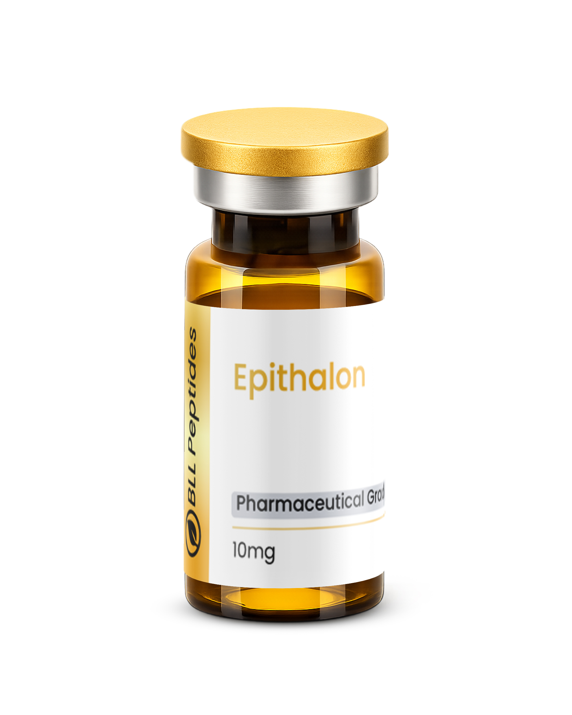
Epithalon (also written Epitalon) is a synthetic tetrapeptide — Ala-Glu-Asp-Gly — developed as a research analog of Epithalamin, a polypeptide extract isolated from bovine pineal gland tissue. The primary mechanism driving scientific interest in Epithalon research is telomerase activation: stimulation of the enzyme responsible for extending and maintaining telomeres, the protective chromosomal caps that shorten with each cell division and correlate strongly with biological aging. Most longevity-focused peptide research operates at the level of growth factor signaling or tissue repair. Epithalon operates at the level of the genome’s clock.
How Epithalon Works: Telomerase, Melatonin, and Antioxidant Defense
As a neurosurgeon who thinks in systems, I was drawn to Epithalon’s multi-layered mechanism profile. Three primary pathways emerge from the research literature:
Telomerase activation: The most mechanistically striking finding in Epithalon research is its ability to activate telomerase — the enzyme that adds nucleotide sequences to telomere ends, counteracting the shortening that accompanies each cell division. In vitro research on human fetal fibroblasts demonstrated that Epithalon could not only activate telomerase but extend the replicative lifespan of cells beyond their typical Hayflick limit. The finding that a four-amino-acid peptide can activate telomerase and extend cellular replicative capacity in vitro is the kind of data that warrants serious mechanistic investigation. Telomere length is now recognized as a robust biomarker of biological age and cumulative cellular stress across multiple tissue types.
Melatonin normalization and pineal regulation: Consistent with its derivation from pineal tissue extract, Epithalon has been shown in animal models to restore nocturnal melatonin secretion — particularly in aged subjects where the pineal gland’s melatonin output has declined. In older rats, Epithalon administration was associated with restoration of nocturnal melatonin peaks toward younger-animal patterns. Melatonin is not merely a sleep hormone; it is a potent endogenous antioxidant with critical roles in circadian regulation, immune modulation, and DNA damage response — making its normalization a meaningful endpoint in longevity research.
Antioxidant and DNA protection: Multiple preclinical studies have documented Epithalon’s effects on markers of oxidative stress — including reductions in lipid peroxidation products and increases in antioxidant enzyme activity (superoxide dismutase, glutathione peroxidase). Oxidative DNA damage accelerates telomere shortening and cellular senescence. These antioxidant effects may work synergistically with telomerase activation, addressing both the rate of telomere shortening and the enzyme available to repair it.
What Epithalon Peptide Research Actually Shows
Animal Longevity Studies
The most substantial data in Epithalon research comes from animal longevity models. In multiple rodent and Drosophila melanogaster model systems, Epithalon treatment was associated with statistically significant increases in both mean and maximum lifespan. A landmark study by Anisimov and colleagues documented lifespan extensions in the range of 24–36% in treated animals versus controls, alongside reduced tumor incidence — a dual-endpoint finding that attracted considerable attention in gerontology. PubMed-indexed research by Anisimov et al. on Epitalon’s effects on lifespan and tumor incidence established the preclinical foundation that the field continues to build on.
Cellular Replicative Lifespan and the Hayflick Limit
The in vitro telomerase work is among the most discussed findings in Epithalon research. Human fetal fibroblast cultures treated with Epithalon showed activation of telomerase and continued cell division beyond the Hayflick limit — the approximately 50–70 divisions normal human somatic cells undergo before entering senescence. This finding does not translate directly to in vivo claims, but it provides a clear mechanistic anchor for the longevity effects observed in animal models and makes Epithalon a valuable tool in cellular aging research.
Immune Aging and Anti-Tumor Research
Research has also documented immune effects consistent with an anti-aging profile: restoration of T-cell function, normalization of interleukin signaling, and improvement in NK cell activity in aged animal models. In cancer model research, Epithalon showed antineoplastic activity — reducing tumor incidence and growth in carcinogen-exposed animals by approximately 27–50% across multiple protocols. This is mechanistically connected to the DNA protection and telomere stabilization data: cells with maintained genome integrity are less susceptible to the mutations that initiate malignant transformation.
Key Findings from Epithalon Peptide Research
- Activates telomerase in human fetal fibroblast cell cultures, extending replicative lifespan beyond the Hayflick limit — the core cellular aging finding
- Produces 24–36% lifespan extension in rodent longevity studies (mean and maximum lifespan) across multiple independent research groups
- Restores nocturnal melatonin secretion patterns in aged animals toward younger-animal baselines, consistent with pineal gland modulation
- Reduces oxidative stress markers including lipid peroxidation; elevates antioxidant enzyme activity (SOD, GPx) in preclinical models
- Reduces tumor incidence by 27–50% in carcinogen-exposed animal models — a finding mechanistically connected to genomic stability and telomere maintenance
- Normalizes T-cell function and NK cell cytotoxic activity in aged animal immune studies
A peptide that simultaneously activates telomerase, normalizes the aging pineal gland’s melatonin output, and reduces tumor incidence in carcinogen models represents one of the most mechanistically coherent longevity research compounds I’ve encountered in this literature.
Epithalon and Complementary Research at BLL Peptides
Researchers exploring the biology of cellular aging and longevity frequently examine Epithalon alongside mechanistically complementary compounds. Our NAD+ research compound addresses the sirtuin pathway and mitochondrial biogenesis — both NAD+ and Epithalon research target upstream biology of cellular aging through distinct but potentially synergistic mechanisms, with NAD+ operating on metabolic and epigenetic aging and Epithalon on the genomic clock itself. For researchers interested in tissue regeneration and growth factor signaling as it intersects with recovery and repair biology, BPC-157 provides a useful mechanistic contrast: where Epithalon targets telomere biology and pineal regulation, BPC-157 operates at the level of angiogenesis, FAK-paxillin signaling, and musculoskeletal repair.
This content is intended for research purposes only. BLL Peptides products are not intended for human consumption.
Frequently Asked Questions About Epithalon
What is Epithalon made of?
Epithalon is a synthetic tetrapeptide consisting of four amino acids: Alanine, Glutamic acid, Aspartic acid, and Glycine (Ala-Glu-Asp-Gly). It was developed as a synthetic analog of Epithalamin — a polypeptide extract from bovine pineal gland tissue — by Professor Vladimir Khavinson and colleagues at the St. Petersburg Institute of Bioregulation and Gerontology in Russia.
How does Epithalon activate telomerase?
In vitro research on human fetal fibroblasts documented that Epithalon upregulates telomerase activity — the enzyme that adds protective nucleotide sequences to telomere ends, counteracting the shortening that accompanies normal cell division. The precise receptor-level mechanism continues to be investigated; current research suggests it may involve epigenetic modulation of telomerase gene expression, including effects on the hTERT (human telomerase reverse transcriptase) promoter region.
What is the Hayflick limit and why does it matter for Epithalon research?
The Hayflick limit is the finite number of times a normal human somatic cell can divide — typically 50–70 divisions in culture — before entering cellular senescence. This limit is tied to telomere shortening: each division removes some telomere sequence, and when telomeres reach critically short length, cell division halts. In vitro research found that Epithalon extended replicative lifespan of human fetal fibroblasts beyond this typical limit through telomerase activation — making the Hayflick limit directly relevant to understanding this research compound’s significance.
Is Epithalon related to melatonin?
Epithalon is not melatonin, but its parent compound Epithalamin was derived from pineal gland tissue — the same gland that produces melatonin. Research has shown that Epithalon administration normalizes melatonin secretion in aged animals, suggesting it modulates pineal gland function rather than directly substituting for melatonin. This pineal-regulatory effect may contribute to Epithalon’s documented antioxidant and immune-aging findings, given melatonin’s well-established roles in DNA damage response and circadian immune regulation.
What animal models have been used in Epithalon longevity research?
Epithalon longevity research has used multiple model systems: rodent models (mice and rats) for lifespan extension and tumor incidence studies, Drosophila melanogaster (fruit fly) models for maximum lifespan endpoints, in vitro human fetal fibroblast cultures for telomerase and Hayflick limit studies, and aged rodent immune models for T-cell and NK cell function endpoints. Khavinson’s research group has also published observational data from human applications in Russian clinical settings, though this represents a much smaller body of evidence than the preclinical literature.
About the Author
Dr. James is a board-certified neurosurgeon and member of the BLL Peptides research advisory team. With decades of surgical experience and a focused interest in neural recovery, neuroprotection, and the emerging science of signaling peptides, Dr. James regularly reviews preclinical literature and translates complex findings for clinicians and researchers working at the frontier of neuroscience.
This content is intended for research purposes only. BLL Peptides products are not intended for human consumption.